Lateralization
Main article: Lateralization of brain function
Routing of neural signals from the two eyes to the brain
Each hemisphere of the brain interacts primarily with one half of the body, but for reasons that are unclear, the connections are crossed: the left side of the brain interacts with the right side of the body, and vice versa.
[citation needed] Motor connections from the brain to the spinal cord, and sensory connections from the spinal cord to the brain, both cross the midline at brainstem levels. Visual input follows a more complex rule: the optic nerves from the two eyes come together at a point called the optic chiasm, and half of the fibers from each nerve split off to join the other. The result is that connections from the left half of the retina, in both eyes, go to the left side of the brain, whereas connections from the right half of the retina go to the right side of the brain. Because each half of the retina receives light coming from the opposite half of the visual field, the functional consequence is that visual input from the left side of the world goes to the right side of the brain, and vice versa. Thus, the right side of the brain receives somatosensory input from the left side of the body, and visual input from the left side of the visual field—an arrangement that presumably is helpful for visuomotor coordination.

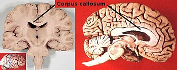
The corpus callosum, a nerve bundle connecting the two cerebral hemispheres, with the lateral ventricles directly below
The two cerebral hemispheres are connected by a very large nerve bundle called the corpus callosum, which crosses the midline above the level of the thalamus. There are also two much smaller connections, the anterior commissure and hippocampal commissure, as well as many subcortical connections that cross the midline. The corpus callosum is the main avenue of communication between the two hemispheres, though. It connects each point on the cortex to the mirror-image point in the opposite hemisphere, and also connects to functionally related points in different cortical areas.
No comments:
Post a Comment